
What is Brain Age?
Brain Age is the biological clock of the brain and represents the aging degree of the brain. It is not equivalent to a person’s chronological age. For example, two people have the same date of birthday, but one person’s Brain Age is older than his/her chronological age, which means that his/her brain is older. And if another person’s Brain Age is younger than his/her chronological age, it means that his/her brain is younger.
How to calculate Brain Age?
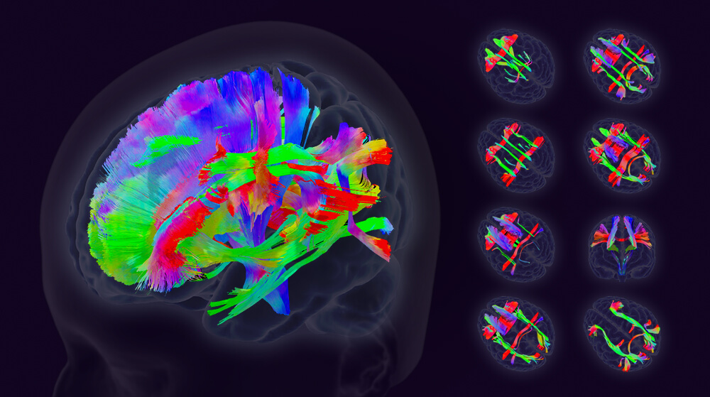
Brain Age is calculated based on changes in different physiological parameters with age. The most common approach is to collect brain images of healthy people across generations and capture brain image features, such as cerebral cortex thickness, brain gray and white matter volume, or nerve fiber tract structure. After marking these features with chronological age, they are input into the artificial intelligence algorithm for training, and finally a statistical prediction model for Brain Age is obtained. This model can predict a person’s Brain Age based on his/her brain images.
Why is Brain Age important?
Recent research shows the importance of Brain Age.
- Brain Age represents a health indicator that combines a person’s innate and acquired factors: Brain Age is affected by many innate factors (such as dementia genes, gender) and acquired factors (such as neurological or mental diseases, metabolic diseases, cardiovascular diseases, or lifestyle factors). Even if two people are born at the same age, their Brain Ages will be different due to different innate and acquired factors.
- Brain Age predicts the risk of future dementia or death: Numerous studies have shown that the above risk factors are closely related to dementia or death, and Brain Age represents a person’s health indicator that combines innate and acquired factors. Recent research has even found that Brain Age predicts future risk of dementia or death. Therefore, Brain Age can predict how close a person is to dementia or death.
- Brain Age can be reversed: Since Brain Age is controlled by many acquired factors, we can keep the brain younger by controlling acquired factors, such as controlling high blood pressure, regular exercise, healthy diet, and adequate sleep. Once the Brain Age is restored to a young age, the risk of dementia or death will be far away, thereby achieving the effect of healthy and long life.
What are the advantages of AcroViz Brain Age technology?
AcroViz combines advanced techniques to develop superior axonal Brain Age technology.
- Excellent stability: AcroViz has a unique image registration technology that can perform a fully automatic image processing program on diffusion magnetic resonance images, and then calculate the Brain Age with excellent stability.
- Higher accuracy: Compared with most gray matter Brain Age estimates using T1-weighted structural images, diffusion MRI is more sensitive to changes in subtle structures, so the estimation of axonal Brain Age is more accurate. For example, the accuracy of AcroViz axonal Brain Age is about 4 to 5 years old. Other research teams used more than three times the amount of T1-weighted structural imaging data to calculate the gray matter Brain Age with an accuracy of about 5 to 6 years.
- Unique network Brain Age: In addition to providing the axonal Brain Age of the entire brain, AcroViz also provides the axonal Brain Age of eight cognitive function networks based on the company’s unique brain fiber tract atlas. These eight cognitive function networks include four memory networks (episodic memory, semantic memory, implicit memory, and working memory) and two high-order functions (language and attention), and two receptive/responsive functions (sensorimotor and visual). The Brain Age of these cognitive function networks can show different degrees of aging among the eight networks.
- Keen clinical relevance: The AcroViz axonal Brain Age focuses more on the degeneration of neuronal connections, while the general gray matter Brain Age focuses more on the degeneration of neuronal bodies. Research shows that the degree of axonal brain aging is more sensitive to clinical symptoms or cognitive function than gray matter Brain Age.
AcroViz Brain Age in clinical research
1. Alzheimer’s disease [1]:
AcroViz used the public database of dementia patients in the United States (OASIS-3) to conduct a retrospective Brain Age study. We found that the older the brain, the worse the clinical dementia rating (CDR). Among patients with mild cognitive impairment, those who progressed to dementia within the next two years had significantly older Brain Ages than those who did not progress. Even among people with normal cognitive function, those who developed mild cognitive impairment within the next two years had older brains than those who did not. Our results show that Brain Age can be used to measure the severity of dementia in a person over 65 years of age. More importantly, Brain Age can predict a normal person’s risk of developing mild cognitive impairment in the future. It can also predict the risk of a patient with mild cognitive impairment deteriorating into dementia in the future.
2. Parkinson’s disease [2,3]:
AcroViz cooperated with the Parkinson’s disease team of National Taiwan University Hospital to conduct Brain Age research, and was the first to publish the manifestation of Brain Age in Parkinson’s disease. We found that Brain Age in Parkinson’s disease was significantly younger than in multiple system atrophy, a subtype of Parkinsonism. We also found that patients with Parkinson’s disease with cognitive impairment had significantly older Brain Age than Parkinson’s disease patients without cognitive impairment or healthy controls. On the contrary, the Brain Age of patients with Parkinson’s disease without cognitive impairment was slightly older than that of healthy people, but the difference was not significant. When we did a correlation analysis between the Brain Age of patients with Parkinson’s disease and their motor or cognitive functions, we found that there was a significant correlation between Brain Age and motor or cognitive functions. The older the Brain Age, the worse the motor or cognitive function scores.
3. Schizophrenia [4]:
AcroViz cooperated with the Schizophrenia Team of National Taiwan University Hospital to conduct Brain Age research. We found that compared with healthy people, the Brain Age of patients with schizophrenia showed significant aging in both gray matter Brain Age and axonal Brain Age. When we did a correlation analysis between Brain Age of patients with schizophrenia and their clinical behavior, we found that there was no correlation between gray matter Brain Age and clinical behavior. However, axonal Brain Age had a significant correlation with age of onset, negative symptoms, and IQ. Patients with older axonal Brain Age had younger onset age, more severe negative symptoms, and lower IQ scores.
4. Mild cognitive impairment [5]:
AcriViz cooperated with the neurology team of Shuang Ho Hospital to conduct a retrospective Brain Age study on patients with mild cognitive impairment taking cholinesterase inhibitors. We found that a patient’s gray matter Brain Age before taking the drug predicted cognitive outcome two years after taking the drug. We divided them into two groups based on their cognitive outcomes after taking the drug for two years. Those whose cognitive function improved were classified into the responsive group, and those whose cognitive function did not improve were classified into the non-responsive group.The results showed that the Brain Age of the responsive group was significantly younger than that of the non-responsive group. This study implies that clinicians can conduct Brain Age tests to predict the prognosis of treatment before administering drugs to patients with mild cognitive impairment, which will help doctors plan for patient treatment.
5. Sub-clinical adults [6]:
AcroViz cooperated with the Institute of Medical Devices and Medical Imaging at the National Taiwan University School of Medicine to use a public aging research database (CamCAN) in the United States to investigate the relationships among cardiovascular health factors, Brain Age, and cognitive functions. We used the statistical mediation method to analyze the causal relationship among the three, and found that Brain Age was the mediator between cardiovascular health factors and cognitive function. Cardiovascular health factors and cognitive function formed a significant correlation through Brain Age. In other words, cardiovascular health factors first affected Brain Age, and Brain Age then affected cognitive function. This research supports improving Brain Age by regulating cardiovascular health factors, thereby achieving the goal of promoting cognitive function.
6. Diffusion magnetic resonance image registration technology [7]:
Although diffusion magnetic resonance imaging can display the microstructural characteristics of brain white matter nerve fibers, such images are easily interfered by a variety of artifacts, including deformation artifacts induced by magnetic susceptibility, deformation artifacts induced by eddy currents, head motion artifacts, etc. These artifacts will seriously affect the calculation results of the microstructural characteristics of nerve fibers, and will also affect the accuracy of the age of nerve fibers. To remove these artifacts, we develop the DACO algorithm. This method uses clinically commonly used T1-weighted structural images as alignment targets, combined with advanced alignment algorithms, to effectively remove artifacts from diffusion magnetic resonance imaging. The DACO algorithm greatly improves the accuracy and stability of Brain Age.
Cooperation projects with hospitals or research institutions
In order to expand the application and clinical value of Brain Age, we have carried out various industry-university cooperation projects with various domestic medical centers, universities or research institutions.
(1) National Taiwan University Hospital: Brain Age Model Establishment (Completed)
(2) Wanfang Hospital: Brain Age Model Establishment (Completed)
(3) Wanfang Hospital: Brain Age Assessment of Dementia Severity (Ongoing)
(4) Shuang Ho Hospital: Prognostic Assessment of Medication Effects for Mild Cognitive Impairment (Completed)
(5) Shuang Ho Hospital: Prognostic Assessment of Stroke Thrombectomy Treatment (Ongoing)
(6) National Institute of Health: Prognostic Assessment of Vascular Dementia (Ongoing)
(7) Chang Gung Memorial University: Prognostic Assessment of Cognitive Function in Breast Cancer (Completed)
(8) Min Sheng General Hospital: Brain Age Model Establishment (Completed)
(9) National Taiwan University: Cognitive Screening Test (Completed)
(10) National Taiwan Normal University: Brain-Physical Fitness Course (Completed)
(11) Cheng Hsin Hospital: Brain Age Model Establishment (Completed)
(12) Kao-Ho Hospital: Brain Age Model Establishment (Completed)
(13) Hualien Tzu Chi Hospital: Brain Age Model Establishment (Ongoing)
(14) Far Eastern Memorial Hospital: Brain Age Assessment of Dementia Severity (Ongoing)
AcroViz Brain Age patents
We have applied Brain Age technology to the analysis of various clinical diseases and obtained US patents. Among them, the first patent has also been applied for patents in the European Union, Canada, Australia, Japan, South Korea, China and other countries.
(1) Using Brain Age to assess current and future cognitive impairment (US11589800B2)
(2) Using Brain Age to assess current and future cognitive status (US11751798B2)
(3) Using Brain Age to predict the prognosis of cognitive impairment treatment (US11883184B1)
(4) Using Brain Age to determine current and future brain atrophy status (US12133739B2)
AcroViz Brain Age papers (Selected)
[1] Tseng, W.Y.I., Hsu, Y.C., Kao, T.W. Brain Age difference at baseline predicts CDR change in approximately two years. J Alzheimer’s Disease. 2022;86(2):613-627.
[2] Chen, C.L., Kuo, M.C., Wu, W.C., Hsu, Y.C., Wu, R.M., Tseng, W.Y.I. Advanced brain aging in multiple system atrophy compared to Parkinson’s disease. Neuroimage Clinical. 2022;34:102997.
[3] Chen, C.L., Cheng, S.Y., Montaser-Kouhsari, L., Wu, W.C., Hsu, Y.C., Tai, C.H., Tseng, W.Y.I., Kuo, M.C., Wu, R.M. Advanced brain aging in Parkinson’s disease with cognitive impairment: A multimodal MRI study. npj Parkinson Disease. 2024 Mar 16;10(1):62. doi: 10.1038/s41531-024-00673-7.
[4] Chen, C.L., Hwang, T.J., Tung, Y.H., Yang, L.Y., Hsu, Y.C., Liu, C.M., Lin, Y.T., Hsieh, M.H., Liu, C.C., Chien, Y.L., Hwu, H.G., Tseng, W.Y.I. Detection of Advanced Brain Aging in Schizophrenia and its Structural Underpinning by Using Normative Brain Age Metrics. Neuroimage Clinical. 2022;34:103003.
[5] Tseng, W.Y.I., Hsu, Y.C., Huang, L.K., Hong, C.T., Lu, Y.H., Chen, J.H., Fu, C.K., Chan, L. Brain Age is associated with cognitive outcomes of cholinesterase inhibitor treatment in patients with mild cognitive impairment. J Alzheimer’s Disease. 2024;98(3):1095-1106. doi: 10.3233/JAD-231109.
[6] Chen, C.L., Kuo, M.C., Chen, P.Y., Tung, Y.H., Hsu, Y.C., Huang, C.W.C., Chan, W.P., and Tseng., W.Y.I. Validation of neuroimaging-based Brain Age gap as a mediator between modifiable risk factors and cognition. Neurobiology of Aging. 2022;114:61-72. [7] Hsu, Y.C., Tseng, W.Y.I. DACO: Distortion/artefact correction for diffusion MRI data. NeuroImage. 2022;262:119571.